
Patient Information:
17 years old, male, American
Mechanism of Injury:
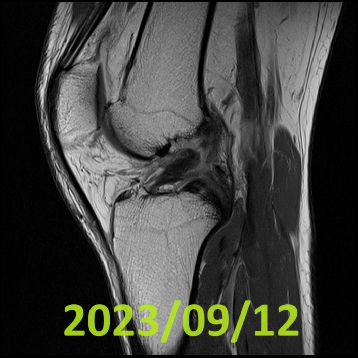
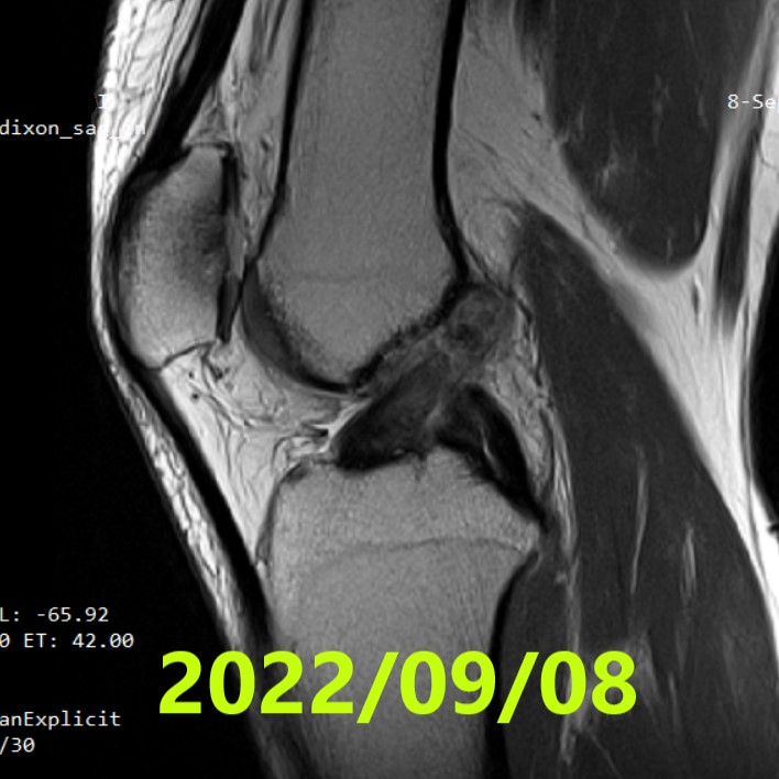
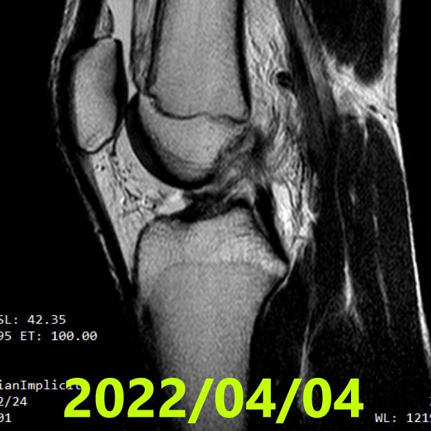
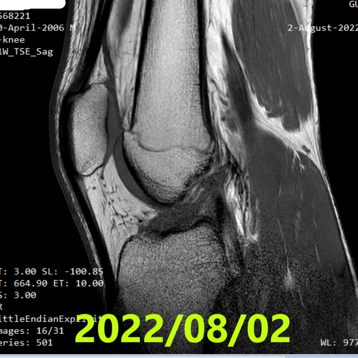
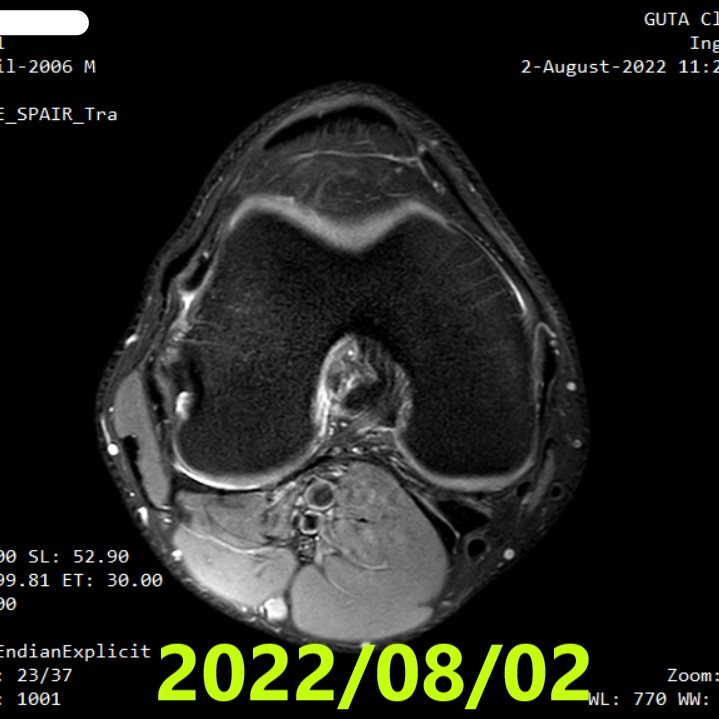
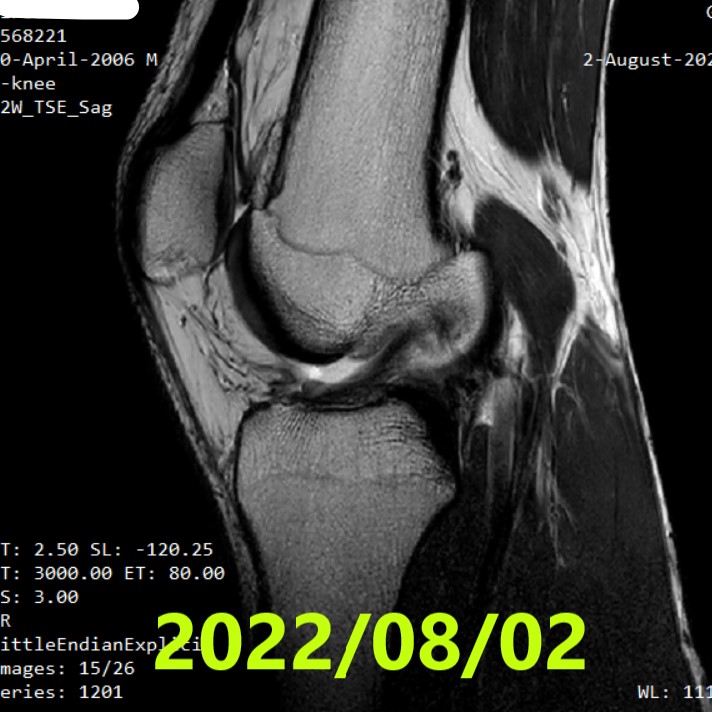
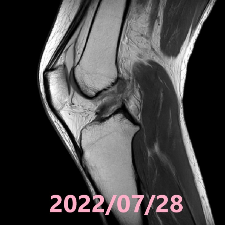
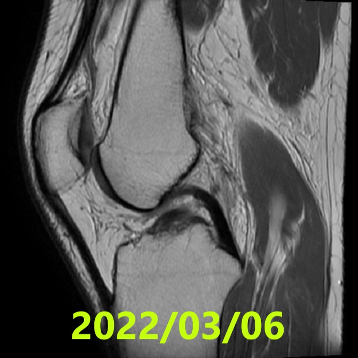
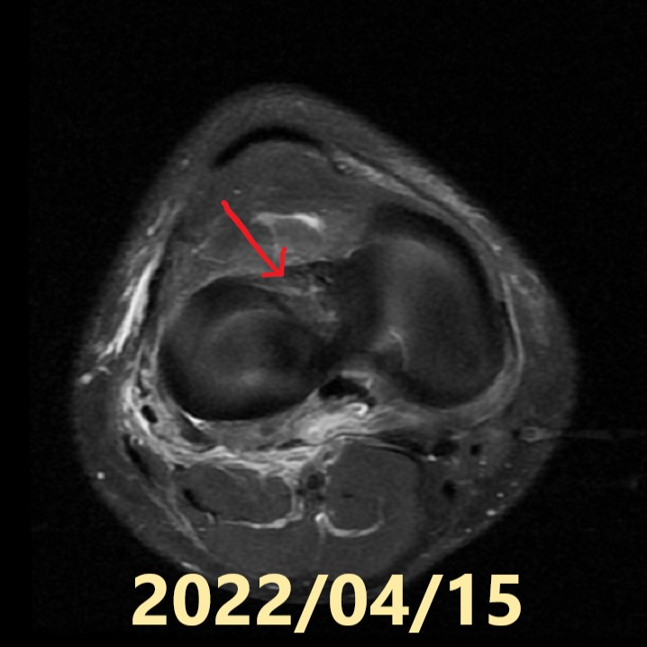
On June 11, 2022, he twisted his right knee after a failed landing from a skateboard jump. An MRI was performed on June 25, and the following diagnosis was obtained:
ACL Tear (unclear tear ends (Ihara Classification IV))
Lateral Collateral Ligament sprain

Post-Injury Progress:
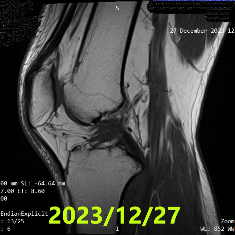
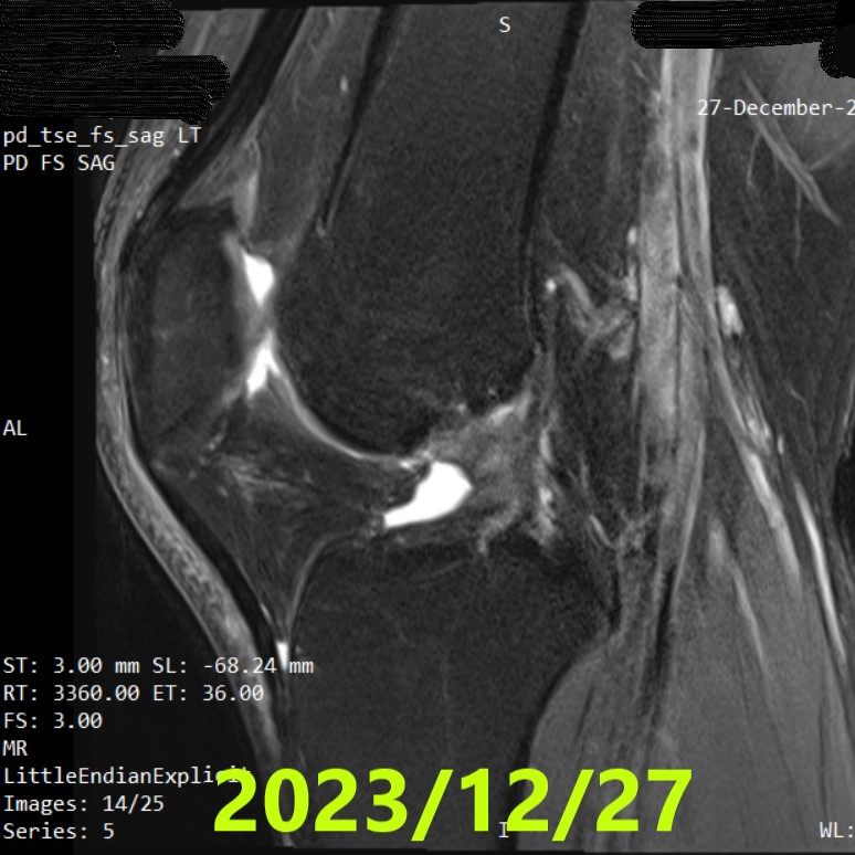
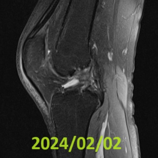
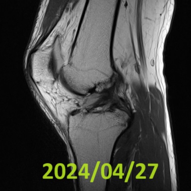
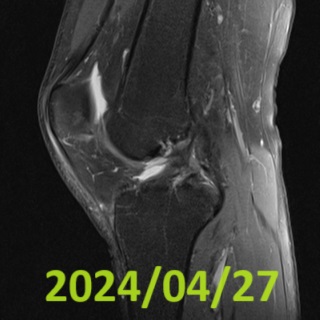
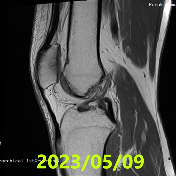
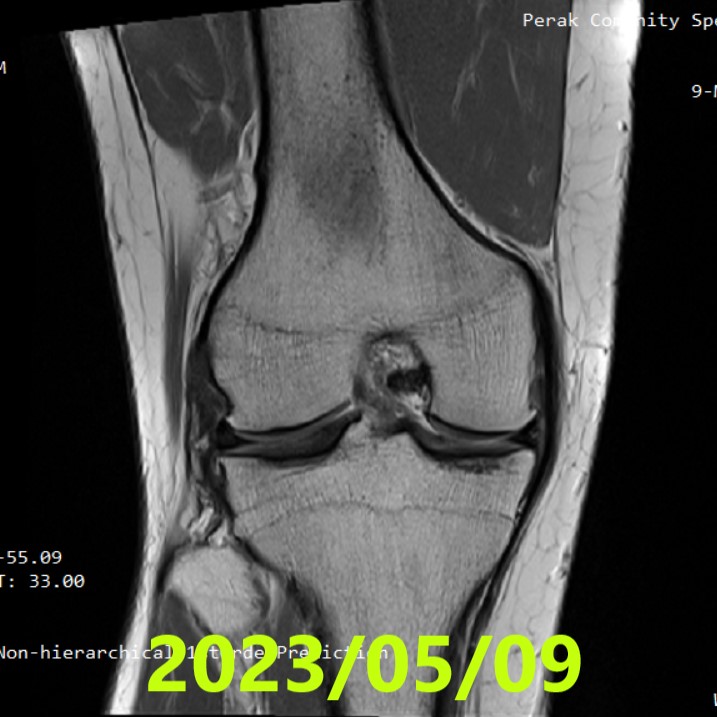
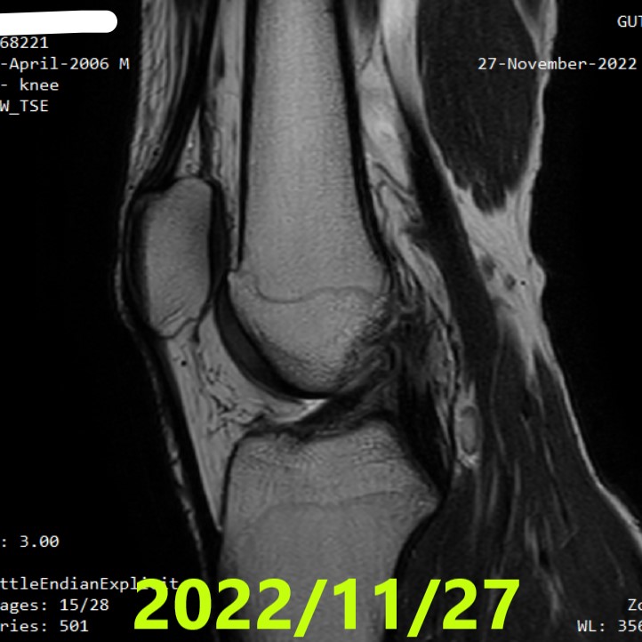
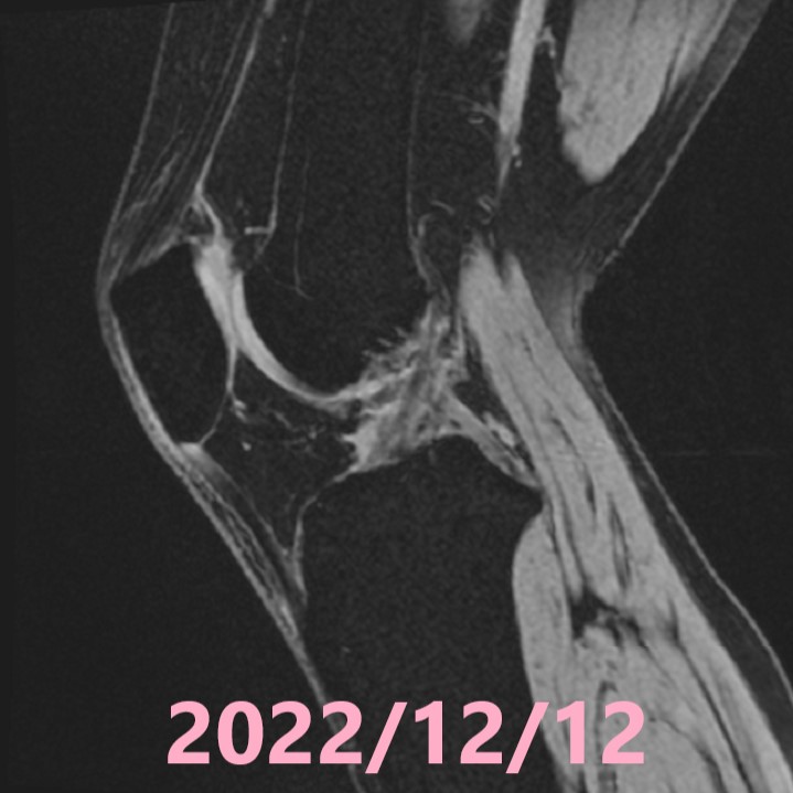
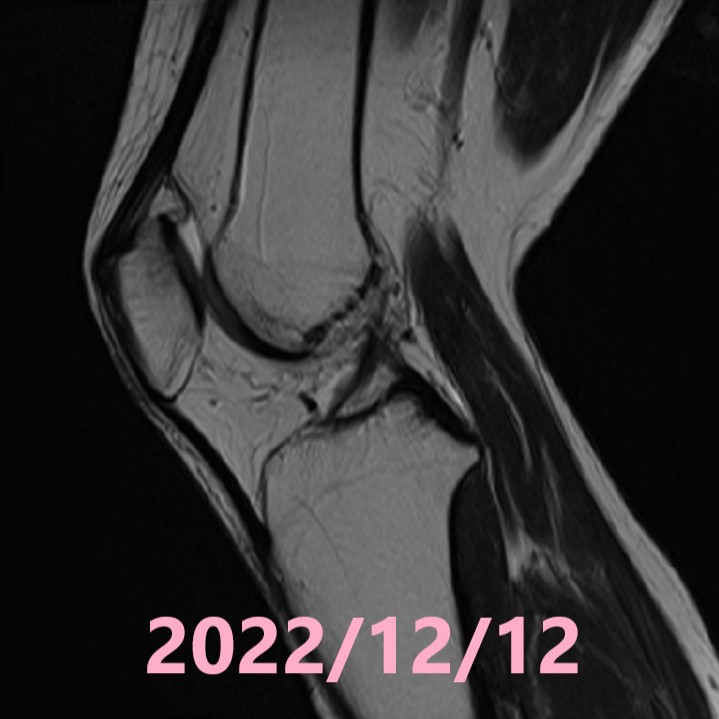
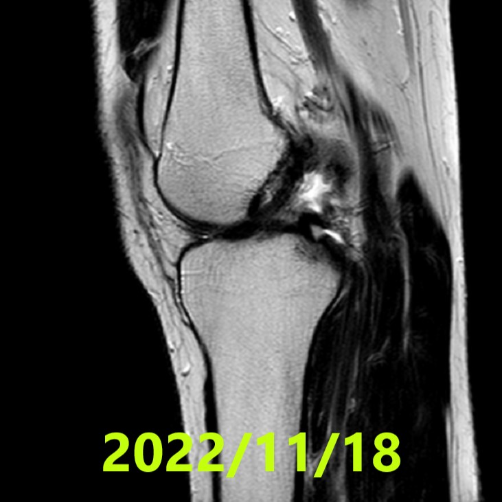
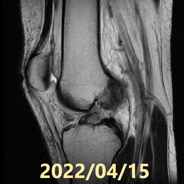
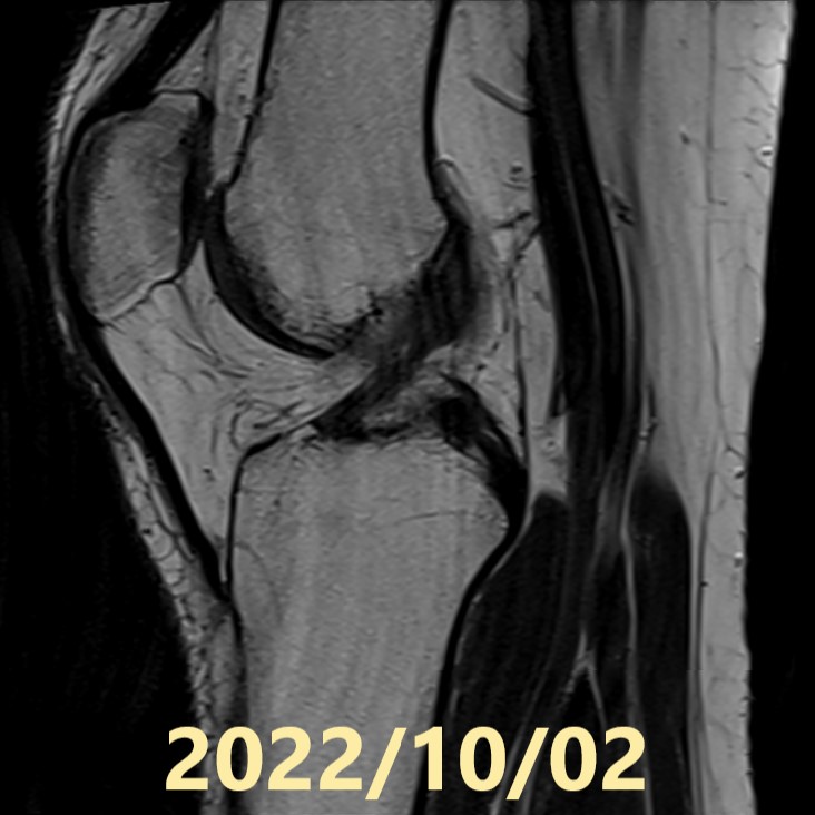
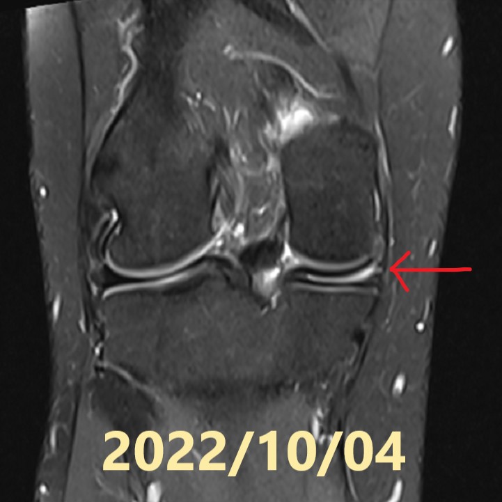
Starting August 3, 2022, he began Jun Matsumoto’s natural therapy through the online sessions. He continued with online treatment and at-home Evo-Devo Exercises, and an MRI was performed five months after the start of treatment. The patient did not use any functional braces.

MRI Results:
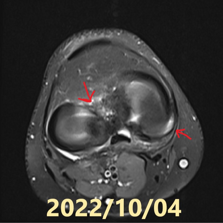
The torn ACL had recovered its continuity with a thick and tense form (Ihara Healing Classification A).
Discussion:
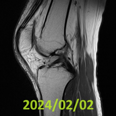
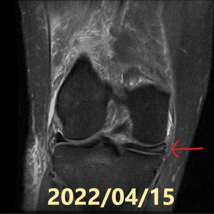
ACL tears in younger individuals often have unclear tear ends due to the softness of their fibers. This case also had unclear tear ends, making natural healing seem difficult. Additionally, the treatment started eight weeks after the injury, which also reduced the likelihood of successful natural healing. However, contrary to expectations, the ACL naturally healed with a thick and tense form. According to the Ihara Healing Classification, this is the highest grade, A. Unfortunately, during the treatment period, the patient sustained mild injuries to the medial and lateral meniscus. The patient was not wearing any functional braces, and I had not instructed him to do so. Considering such cases, starting from 2023, I have instructed all patients to wear braces.
References:
- Ihara H, Miwa M, Deya K, Torisu K. MRI of anterior cruciate ligament healing. J Comput Assist Tomogr. 1996 Mar-Apr;20(2):317-21. doi
- Ihara H, Kawano T. Influence of Age on Healing Capacity of Acute Tears of the Anterior Cruciate Ligament Based on Magnetic Resonance Imaging Assessment. J Comput Assist Tomogr. 2017 Mar/Apr;41(2):206-211. doi
- Pitsillides A, Stasinopoulos D, Giannakou K. Healing potential of the anterior cruciate ligament in terms of fiber continuity after a complete rupture: A systematic review. J Bodyw Mov Ther. 2021 Oct;28:246-254. doi
- Filbay, Stephanie R et al. “Healing of acute anterior cruciate ligament rupture on MRI and outcomes following non-surgical management with the Cross Bracing Protocol.” British journal of sports medicine, bjsports-202