
Injury during a soccer match: the ACL was completely torn, and the medial collateral ligament was partially torn. No surgery was performed. Instead, the patient completed 3 months of online Evo-Devo Exercise ( avoid full knee extension / ≤3,000 steps per day / home exercises 3 times daily ). At 4 months, MRI (2023/8/31) confirmed restored continuity (ACLOAS 2), and this was maintained at 11 months (2024/2/6). Daily activities were problem-free, but the patient had not yet returned to sport.
ACL Online Therapy for natural healing
The case reports of ACL natural healing
Patient Information
- Age / Nationality: 49-year-old male, born in Morocco, American
- Occupation: Construction company employee (heavy work avoided during treatment; on-site walking continued)
- History: Meniscal injury (for ~5 years)
Mechanism of Injury
- Date of injury: 19 March 2023
- Context: Slipped while playing soccer and injured the right knee with flexion and twist
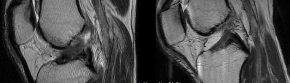
Initial Evaluation (MRI #1)
- Date: 7 April 2023
- Diagnosis: Right knee complete ACL rupture + partial tear of the medial collateral ligament
- Independent readings (three radiologists): unanimous Ihara classification IV (indistinct/poorly formed stumps)
[Ihara classification notes]
I = linear/straight tear; II = curved tear (simple complete rupture); III = tear with displaced stumps; IV = indistinct/unclear stumps (most complex).
Initial Policy & Start of Therapy
- Start date: 22 April 2023
- Format: Online care at Matsumoto Jun clinic; initiation of Evo-Devo Exercise
- Brace management: knee extension limited to 0–30°
- Activity: walking allowed up to ≤3,000 steps/day (on-site patrol/walking continued)
- Home program: Evo-Devo Exercise 3 times/day for 3 months
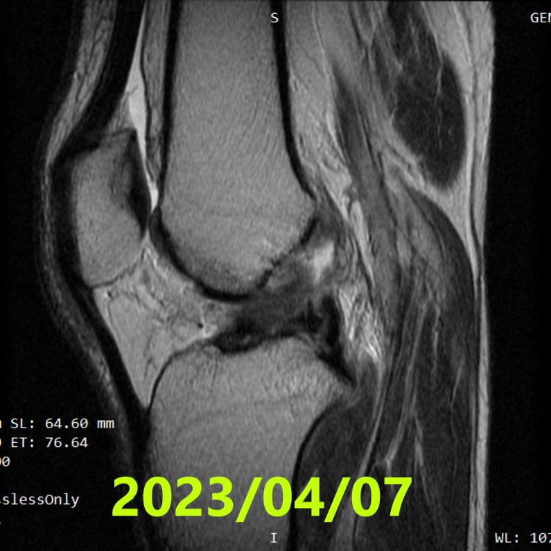
Second MRI (~4 months)
- Date: 31 August 2023
- Independent readings (three radiologists): unanimous ACLOAS grade 2 (thinning/elongation with continuity preserved)
- Interpretation: from complex Ihara IV appearance to findings indicating restored continuity
[ACLOAS (native ACL) notes]
0 = normal (low signal, regular); 1 = thickening/intra-ligament high signal with shape and continuity preserved; 2 = thinning/elongation with continuity preserved; 3 = discontinuity (defect).
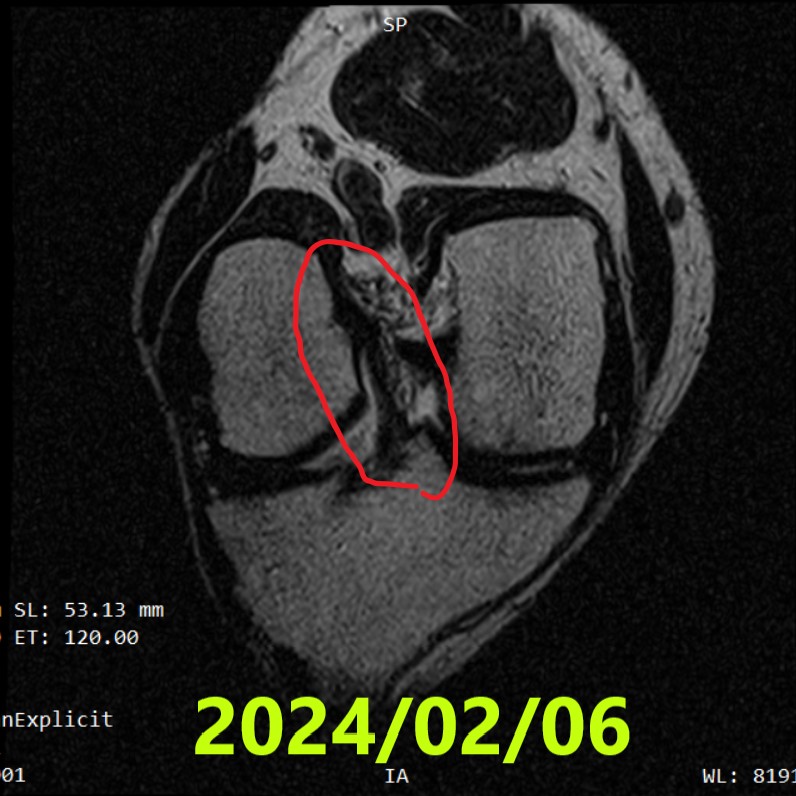
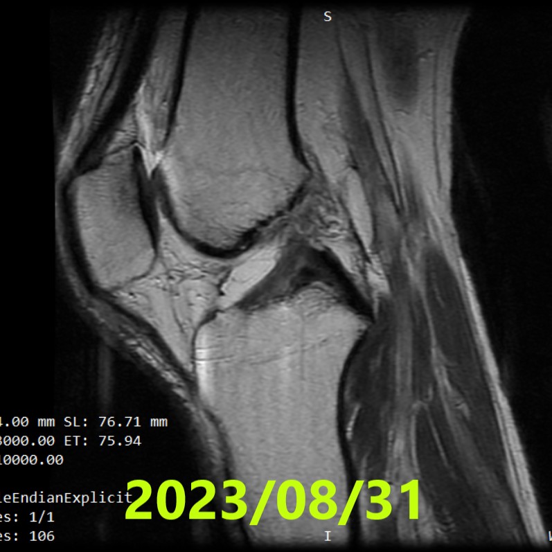
Third MRI (~11 months)
- Date: 6 February 2024
- Findings: no major change, but the ACL’s continuity remained clearly preserved
Subsequent Management (after MRI #2)
- After confirmation of continuity on MRI #2, progressive recovery of full extension and range of motion
- Strengthening: began quadriceps-focused knee strengthening
- Course: by ~11 months after injury, daily activities were performed without problems
- Return to sport: not resumed (patient preference due to age and concern about re-injury)
Results
- Initial: Ihara IV (most complex pattern)
- ~4 months: ACLOAS 2 (continuity restored)
- ~11 months: continuity again confirmed; daily life without problems
Summary
- Injury (2023/03/19): right knee complete ACL rupture + partial tear of the medial collateral ligament (slip/twist while playing soccer)
- MRI #1 (2023/04/07): Ihara IV (unanimous, 3/3 readers)
- Conservative plan: online Evo-Devo Exercise with weekly follow-up + brace limiting extension 0–30° + ≤3,000 steps/day + home exercise 3×/day
- MRI #2 (2023/08/31): ACLOAS 2 (unanimous, 3/3 readers) — continuity restored
- MRI #3 (2024/02/06): continuity preserved; daily activities problem-free (no sport return)
References
- Filbay SR, Dowsett M, Jomaa MC, et al. Healing of acute anterior cruciate ligament rupture on MRI and outcomes following non-surgical management with the Cross Bracing Protocol. Br J Sports Med. 2023;57(23):1490–1497.
- Ihara H, Kawano T. Influence of Age on Healing Capacity of Acute Tears of the ACL Based on MRI Assessment. J Comput Assist Tomogr. 2017;41(2):206–211.
- Roemer FW, Frobell R, Lohmander LS, et al. Anterior Cruciate Ligament OsteoArthritis Score (ACLOAS): Longitudinal MRI-based whole joint assessment of ACL injury. Osteoarthritis Cartilage. 2014;22(5):668–682.
Leave a Reply