
ACL Natural Healing Case Report #50: Most Severe Ihara Type IV Successfully Regenerated to ACLOAS Grade 0
Summary:
Complete ACL rupture suffered during a snowboarding jump landing. Following a strict consensus rule among three independent specialists, the initial diagnosis was Ihara Type IV—the most severe/complex rupture. The patient immediately mastered “full hip external rotation” during online Evo-Devo Exercise guidance. Through rigorous alignment management (0–30° brace / ≤3,000 steps per day) and consistent home exercises 3 times daily, the final MRI achieved the highest possible rating: ACLOAS Grade 0 (regeneration to normal ligament structure). The patient has successfully returned to activities of daily living and phased sports participation.
ACL Online Therapy for natural healing
The case reports of ACL natural healing
Patient Information
- Age / Nationality: 41-year-old male, Japanese
- Injured side: Left knee
Mechanism of Injury
- Date of injury: 1 April 2023
- Context: Sustained a heavy impact during a snowboarding jump landing; an audible “pop” was confirmed at the time of injury
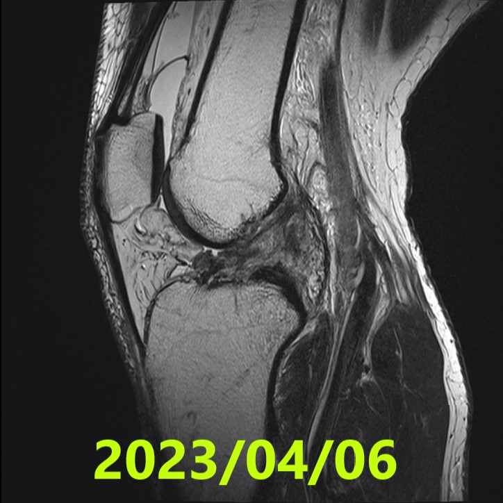
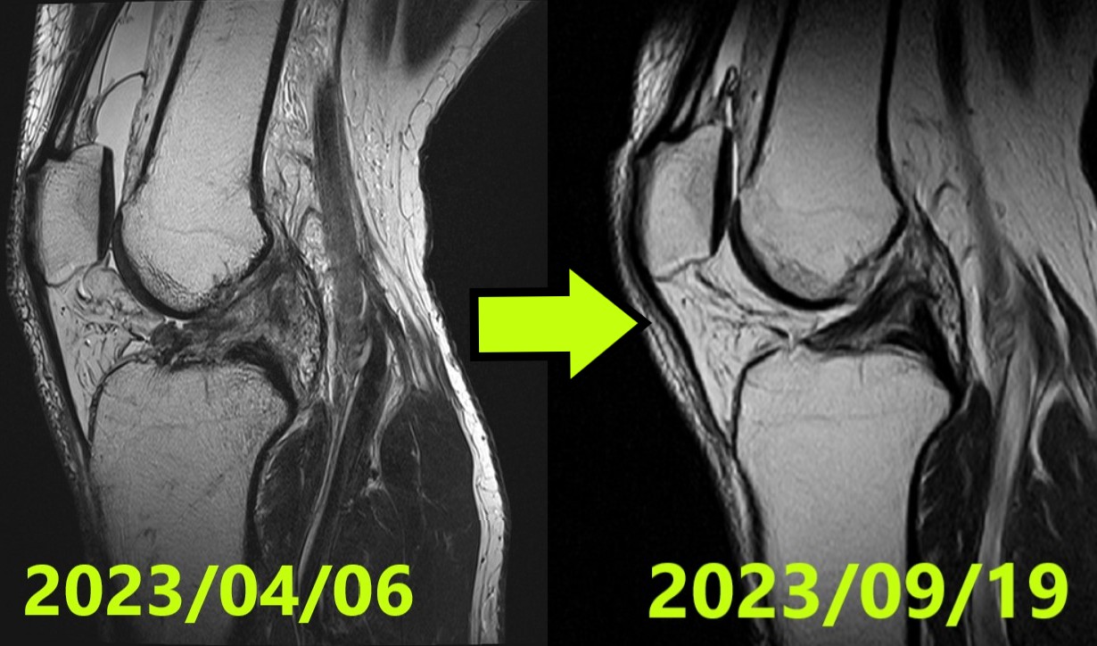
Initial Evaluation (MRI #1)
- Date: 6 April 2023
- Diagnosis: Left knee complete ACL rupture
- Ihara classification: IV (Grade 4) — Confirmed by consensus of 3 independent specialists
- Note: Following the injury, the patient strictly avoided full knee extension and weight-bearing resistance training
[Ihara classification notes]
I = linear/straight tear; II = curved tear (simple complete rupture); III = tear with displaced stumps; IV = indistinct/unclear stumps (most complex).
Initial Policy & Start of Therapy
- Start date: 8 April 2023
- Format: Online care at Matsumoto Jun clinic; initiation of Evo-Devo Exercise
- Brace management: Knee extension strictly limited to 0–30°
- Activity: Walking restricted to ≤3,000 steps/day
- Home program: Evo-Devo Exercise 3 times/day
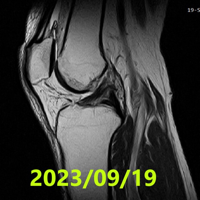
Final MRI Evaluation (~5 months)
- Date: 19 September 2023
- Assessment: ACLOAS Grade 0 (regeneration to normal structure) — Confirmed by consensus of 3 independent specialists
- Finding: The previously ruptured ligament achieved high-quality regeneration with morphology identical to a healthy ligament
[ACLOAS (native ACL) notes]
0 = normal (low signal, regular); 1 = thickening/intra-ligament high signal with shape and continuity preserved; 2 = thinning/elongation with continuity preserved; 3 = discontinuity (defect).
Author’s Observations
- Achieving ACLOAS Grade 0 from the initial “Ihara Type IV” (the most severe classification) is a remarkable clinical milestone
- The patient demonstrated exceptional mastery of hip external rotation (perfect execution of the “Frog Pose”) immediately upon instruction
- This immediate correction of knee alignment likely played a pivotal role in facilitating the highest quality of natural ligament regeneration
Results
- Initial: Ihara IV (consensus by 3 specialists)
- Final: ACLOAS Grade 0 (consensus by 3 specialists) — restored normal ligament structure
- Clinical status: Full return to ADL; currently undergoing phased sports rehabilitation
Summary
- Injury (2023/04/01): Left knee complete ACL rupture (snowboarding jump landing)
- MRI #1 (2023/04/06): Ihara IV — confirmed as severe complete rupture by 3 specialists
- Evo-Devo Plan: Online guidance + strict 0–30° bracing + ≤3,000 steps/day + mastery of hip external rotation
- MRI #2 (2023/09/19): ACLOAS 0 — confirmed as high-quality regeneration to normal structure by 3 specialists
- Outcome: Precise alignment management allowed even a Type IV rupture to achieve the highest grade of natural healing
References
- Filbay SR, Dowsett M, Jomaa MC, et al. Healing of acute anterior cruciate ligament rupture on MRI and outcomes following non-surgical management with the Cross Bracing Protocol. Br J Sports Med. 2023;57(23):1490–1497.
- Ihara H, Kawano T. Influence of Age on Healing capacity of Acute Tears of the ACL Based on MRI Assessment. J Comput Assist Tomogr. 2017;41(2):206–211.
- Roemer FW, Frobell R, Lohmander LS, et al. Anterior Cruciate Ligament OsteoArthritis Score (ACLOAS): Longitudinal MRI-based whole joint assessment of ACL injury. Osteoarthritis Cartilage. 2014;22(5):668–682.
Leave a Reply