Injury while dancing: the ACL was completely torn, and the medial collateral ligament was partially torn. No surgery was performed. Instead, the patient completed 3 months of online Evo-Devo Exercise ( avoid full knee extension / ≤3,000 steps per day / home exercises 3 times daily ). At ~4 months, MRI (2023/8/27) confirmed restored continuity (ACLOAS 2). At ~21 months (2025/1/7), MRI suggested further healing progression, but it did not reach ACLOAS 1. Daily activities were problem-free; return to sport has not yet been confirmed.
ACL Online Therapy for natural healing
The case reports of ACL natural healing
Patient Information
- Age / Nationality: 25-year-old female, Lithuanian
Mechanism of Injury
- Date of injury: 1 April 2023
- Context: While dancing, she missed a floor level change/step and twisted the right knee
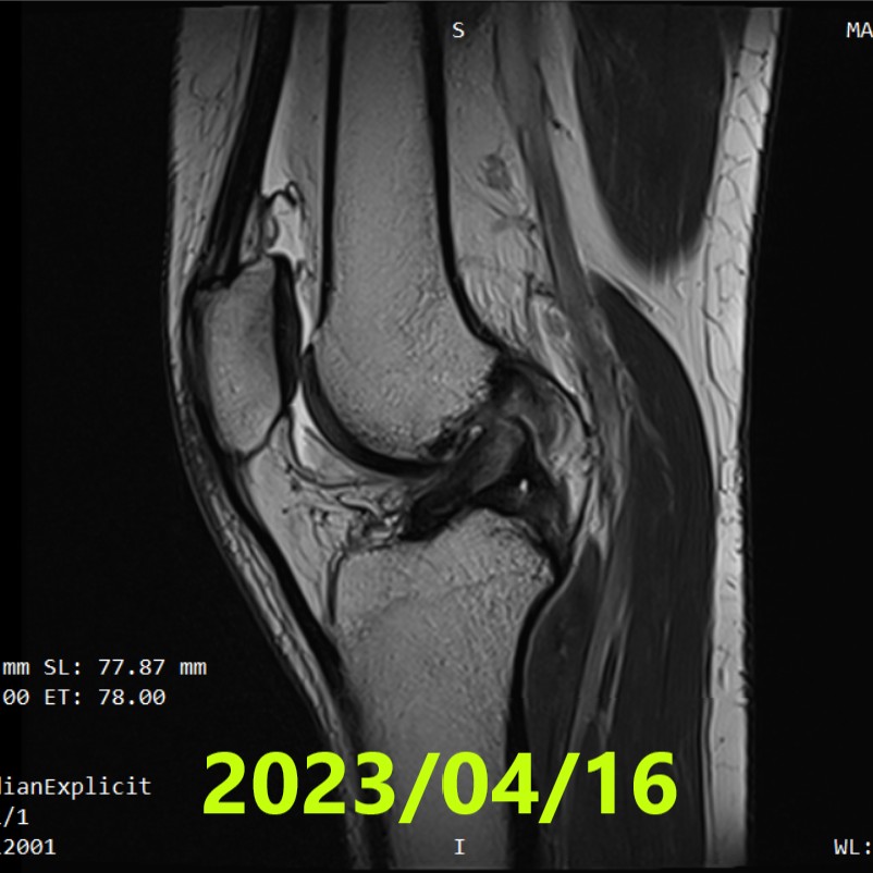
Initial Evaluation (MRI #1)
- Date: 16 April 2023
- Diagnosis: Right knee complete ACL rupture + partial tear of the medial collateral ligament
- Tear pattern: Proximal tear at the femoral attachment (proximal one-third)
- Ihara classification: II (Grade 2)
- Note: No physiotherapy was performed. Full knee extension was performed only during the MRI examination
[Ihara classification notes]
I = linear/straight tear; II = curved tear (simple complete rupture); III = tear with displaced stumps; IV = indistinct/unclear stumps (most complex).
Initial Policy & Start of Therapy
- Start date: 27 April 2023
- Format: Online care at Matsumoto Jun clinic; initiation of Evo-Devo Exercise
- Brace management: knee extension limited to 0–30° (flexion beyond 30° was unrestricted)
- Activity: walking allowed up to ≤3,000 steps/day
- Home program: Evo-Devo Exercise 3 times/day for 3 months
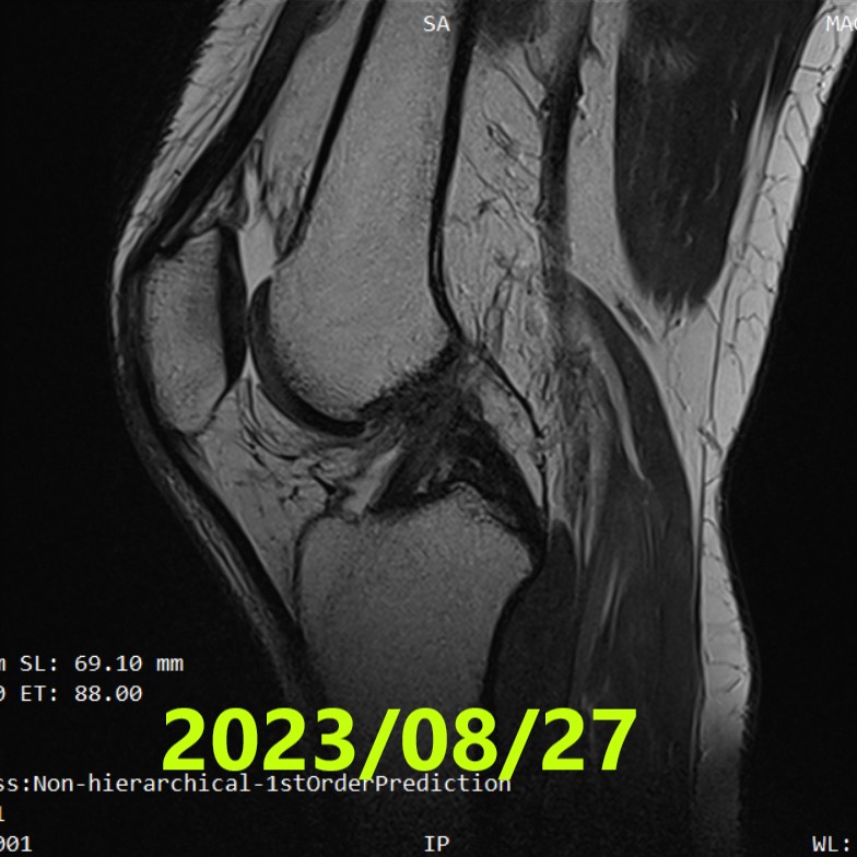
Second MRI (~4 months)
- Date: 27 August 2023
- Assessment: ACLOAS grade 2 (thinning/elongation with continuity preserved)
- Interpretation: findings indicating restored continuity
[ACLOAS (native ACL) notes]
0 = normal (low signal, regular); 1 = thickening/intra-ligament high signal with shape and continuity preserved; 2 = thinning/elongation with continuity preserved; 3 = discontinuity (defect).
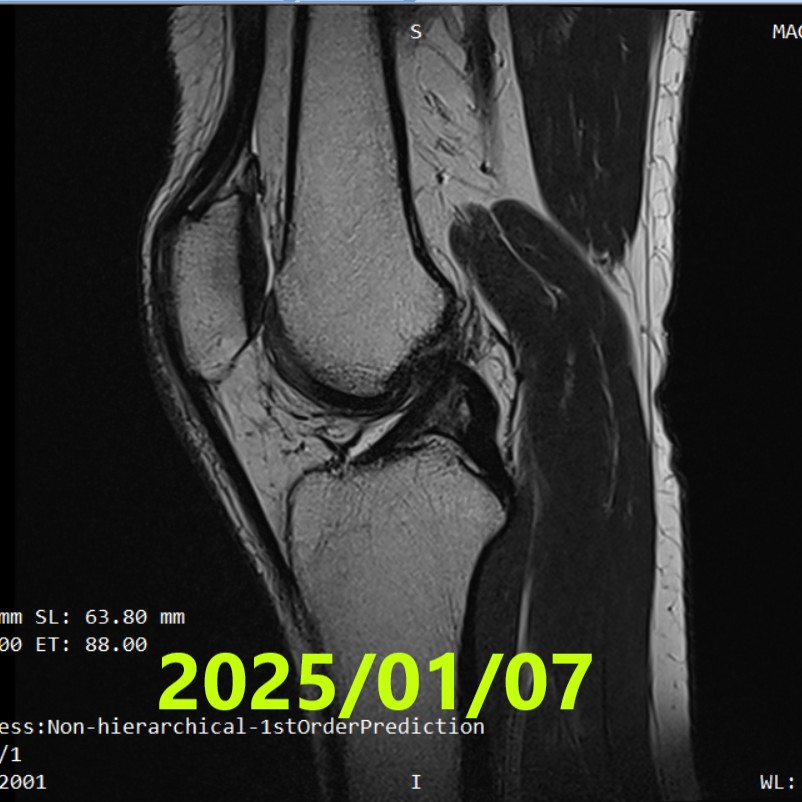
Third MRI (~21 months)
- Date: 7 January 2025
- Findings: Compared with MRI #2, the ACL signal appeared denser, suggesting further healing progression
- Assessment: did not reach ACLOAS grade 1
Subsequent Management (after MRI #2)
- After confirmation of continuity on MRI #2, progressive recovery of full extension and range of motion
- Strengthening: began knee strengthening exercises
- Course: daily activities were performed without problems
- Activity level: cycling and running were performed
- Return to sport: not yet confirmed
Results
- Initial: Ihara II (simple complete rupture pattern) + partial MCL tear
- ~4 months: ACLOAS 2 (continuity restored)
- ~21 months: findings suggesting further healing progression (did not reach ACLOAS 1); daily life without problems
Summary
- Injury (2023/04/01): right knee complete ACL rupture + partial tear of the medial collateral ligament (twist while dancing)
- MRI #1 (2023/04/16): Ihara II (proximal tear at femoral attachment)
- Conservative plan: online Evo-Devo Exercise + brace limiting extension 0–30° + ≤3,000 steps/day + home exercise 3×/day
- MRI #2 (2023/08/27): ACLOAS 2 — continuity restored
- MRI #3 (2025/01/07): suggested further healing progression (did not reach ACLOAS 1)
- After MRI #2: progressive full extension & strengthening; daily activities problem-free (return to sport unconfirmed)
References
- Filbay SR, Dowsett M, Jomaa MC, et al. Healing of acute anterior cruciate ligament rupture on MRI and outcomes following non-surgical management with the Cross Bracing Protocol. Br J Sports Med. 2023;57(23):1490–1497.
- Ihara H, Kawano T. Influence of Age on Healing Capacity of Acute Tears of the ACL Based on MRI Assessment. J Comput Assist Tomogr. 2017;41(2):206–211.
- Roemer FW, Frobell R, Lohmander LS, et al. Anterior Cruciate Ligament OsteoArthritis Score (ACLOAS): Longitudinal MRI-based whole joint assessment of ACL injury. Osteoarthritis Cartilage. 2014;22(5):668–682.